Long Bone Diagram Inside / Skeletal System 1 The Anatomy And Physiology Of Bones Nursing Times - Click on the tags below to find other quizzes on the same subject.
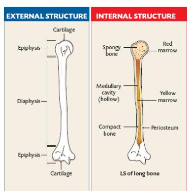
Long Bone Diagram Inside / Skeletal System 1 The Anatomy And Physiology Of Bones Nursing Times - Click on the tags below to find other quizzes on the same subject.. Long bone tissue diagram / introduction to bone boundless anatomy and physiology / a long bone has two main regions:.this is covered by a membrane of connective tissue called the periosteum.beneath the cortical bone layer is a layer of spongy cancellous bone.inside this is the medullary cavity which has an inner core of bone marrow, it contains nutrients and help in formation of cells, made up. A picture of the four bone layers. This is an online quiz called label the long bone. Inside this is a layer of spongy (cancellous) bone which contains red bone marrow. Long bones, especially the femur and tibia, are subjected to most of the load during daily activities and they are crucial for skeletal mobility.
There is a printable worksheet available for download here so you can take the quiz with pen and paper. Newborn babies are actually born with many more bones than this (around 300), but many bones grow together, or fuse, as babies become older. A long bone has two parts: A long bone is a bone that has greater length than width. Long bone diagram inside :
Epiphysis Wikipedia from upload.wikimedia.org A long bone has two parts: A long bone is a bone that has greater length than width. Related posts of inside a bone diagram. Diagram of of a long bone. A long bone is a bone that has greater length than width. This video also explains how damage to epiphyseal artery may lead to avascular necrosis in some. This is an online quiz called label the long bone. The diaphysis and the epiphysis.
The femur or thighbone is the longest and largest bone in the human body.
4 looking at the inside of the bone. The diaphysis is the tubular shaft that runs between the proximal and distal ends of the bone. The diaphysis and the epiphysis (figure 6.3.1). This video also explains how damage to epiphyseal artery may lead to avascular necrosis in some. Gross anatomy of bones a long bone has two main regions: The hollow region in the diaphysis is called the medullary cavity, which is filled with yellow. The blood vessels inside a bone. Besides having a significant length vs width when compared. The outer part of a long bone is made of compact bone. The diaphysis is the tubular shaft that runs between the proximal and distal ends of the bone. Long bone diagram inside : The structure of a long bone allows for the best visualization of all of the parts of a bone ((figure)). The shiny, articulating cartilage on the ends of a bone.
A long bone has two parts: Others are thin, flat, and wide, like your shoulder blades. The hollow region in the diaphysis is called the medullary cavity, which is filled with yellow. The femur or thighbone is the longest and largest bone in the human body. Long bone labeling diagram quizlet from o.quizlet.com bones are also very good at repairing themselves.
Bone Simple English Wikipedia The Free Encyclopedia from upload.wikimedia.org This is an online quiz called label the long bone. Bone is found in the shafts of long bone and consists of various cylindrical units named as haversian system 47. Ends of long bones, pelvic bones, ribs, skull, and vertebrae thin rods and plates of bone tissue. The diaphysis is the tubular shaft that runs between the proximal and distal ends of the bone. Besides having a significant length vs width when compared. Just in case you get tired of looking at the screen we've provided images and pdf files that you can. The diaphysis is the tubular shaft that runs between the proximal and distal ends of the bone. A long bone is one that is cylindrical in shape, being longer than it is wide.keep in mind, however, that the term describes the shape of a bone, not its size.
The diaphysis and the epiphysis.
The hollow region in the diaphysis is called the medullary cavity, which is filled with yellow. The structure of a long bone allows for the best visualization of all of the parts of a bone (). The femur or thighbone is the longest and largest bone in the human body. This diagram will show you. A long bone has two parts: The outer part of a long bone is made of compact bone. A long bone has two parts: A typical long bone shows the gross anatomical characteristics of bone. The shiny, articulating cartilage on the ends of a bone. Added together, your bones make up about 15% of your body weight. The interior part of a long bone consists of the medullary cavity. Long bones include the humerus (upper arm), radius (forearm), ulna (forearm), femur (thigh), fibula (thin bone of the lower leg), tibia (shin bone), phalanges (digital bones in the hands and feet), metacarpals (long bones within the hand), and metatarsals (long bones within the feet). Diagram of inside of skull manual e books.
This is covered by a membrane of connective tissue called the periosteum.beneath the cortical bone layer is a layer of spongy cancellous bone.inside this is the medullary cavity which has an inner core of bone marrow, it contains nutrients and help in formation of cells, made up of yellow marrow in. The tough membrane covering the shaft of the bone. Click on the tags below to find other quizzes on the same subject. The structure of a long bone allows for the best visualization of all of the parts of a bone (). The outer shell of the long bone is made of cortical bone also known as compact bone.
Structure Of A Long Bone Level 2 Anatomy And Physiology from parallelcoaching.co.uk Just in case you get tired of looking at the screen we've provided images and pdf files that you can. A long bone is one that is cylindrical in shape, being longer than it is wide.keep in mind, however, that the term describes the shape of a bone, not its size. The dorsal radial zone is the area of the wrist near the thumb over the palm side. This is an online quiz called long bone anatomy. This is covered by a membrane of connective tissue called the periosteum.beneath the cortical bone layer is a layer of spongy cancellous bone.inside this is the medullary cavity which has an inner core of bone marrow, it contains nutrients and help in formation of cells, made up of yellow marrow in. The blood vessels inside a. The diaphysis is the tubular shaft that runs between the proximal and distal ends of the bone. In a long bone, this is normally found at either end of the bone, in flat or irregular bones it is a thin layer found just inside the compact bone.
A typical long bone shows the gross anatomical characteristics of bone.
The outer shell of the long bone is made of cortical bone also known as compact bone. The tough membrane covering the shaft of the bone. Long bones, especially the femur and tibia, are subjected to most of the load during daily activities and they are crucial for skeletal mobility. A typical long bone shows the gross anatomical characteristics of bone. Add to favorites 0 favs. A long bone has two parts: The diaphysis is the hollow, tubular shaft that runs between the proximal and distal ends of the bone. The structure of a long bone allows for the best visualization of all of the parts of a bone ((figure)). Interestingly, compact bone constitutes up to 80% of the bone's weight, with spongy bone making up the additional 20%, despite its much larger surface area. Long bone labeling diagram quizlet from o.quizlet.com bones are also very good at repairing themselves. The shiny, articulating cartilage on the ends of a bone. Inside the diaphysis is the medullary cavity, which is filled with yellow bone marrow in an adult. A long bone has two parts:
Inside this is a layer of spongy (cancellous) bone which contains red bone marrow long bone diagram. Inside this is a layer of spongy (cancellous) bone which contains red bone marrow.


0 Komentar